Sample Preparation Gallery
From Ug11bm
Jump to navigationJump to search
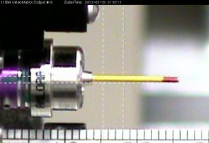
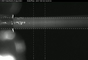
Two views of a sample in position for data-taking, as seen through the "upstream" camera (left) and the video microscope (right). The reticle lines show the nominal extent of the beam. The actual beam edges extend somewhat farther, so these areas should be kept clear of foreign material (glue, glay, wax, etc.).

